What you will learn
- 1Magnification and resolution
- 2The parts of a light microscope and how to prepare a slide
- 3Using I = A × M and converting units
- 4How light and electron microscopes compare
Read
Magnification: How many times larger the image is compared to the actual object.
Resolution: The ability to distinguish two very close points as separate and clear.
01
Light Microscope (Optical Microscope)
- Uses visible light and glass lenses to magnify specimens.
- Can view living cells.
- Maximum useful magnification: ~×1500.
- Resolution: lower than electron microscopes → cannot see very small structures like ribosomes or plasmids.
- lower than electron microscopes → cannot see very small structures like ribosomes or plasmids.
02
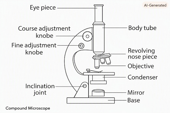
Main parts of light/compound microscopy
- Eyepiece lens – usually ×10.
- Objective lenses – e. g., ×4, ×10, ×40.
- Coarse focus knob – large adjustments.
- Fine focus knob – sharpens image.
- Stage – holds slide.
- Light source / mirror – illuminates’ specimen.
- Iris diaphragm – controls light intensity.
03
Preparing a Slide (Light/compound microscope)
- Specimen must be thin and transparent to allow light through.
- Place specimen in water or saline (NaCl solution) for live cells.
- Add stain if needed (e. g., iodine for plant cells).
- Lower coverslip gently using a mounted needle to avoid air bubbles.
Formula triangle to find magnification/image size/actual size:
- The magnification of an image can be calculated by simple formula.
- Magnification = size of the image / actual size of the object.
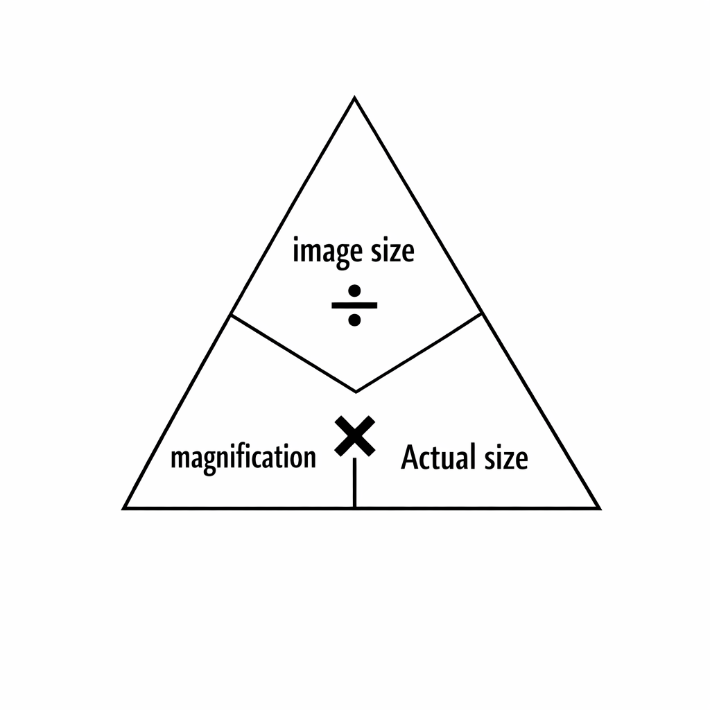
- You must cover the part of the triangle to find and follow the equation that is left behind.
- For example, to find out image size, the formula is magnification x actual size.
- To find out magnification, the formula is image size / actual size.
- To find out actual size of the cell, the formula is image size / magnification.
04
Magnification Formula
- Total magnification = eyepiece × objective.
- For size calculations:
- I = A X M, where:
- I = image size
- A = actual size
- M = magnification
- Rearranged:
05
M = I ÷ A
06
A = I ÷ M
Important: Use the same units for I and A before calculating.
07
Units & Conversions
- Microscopy often uses nm, μm, mm. Conversion chain:
- nm → μm: ÷1000
- μm → mm: ÷1000
- mm → cm: ÷10
- cm → m: ÷100
- Example: 2,500,000 nm → 2500 μm → 2.5 mm → 0.25 cm → 0.0025 m.
08
Electron Microscopes
- Use electrons instead of light → much higher magnification and resolution.
- Types:
- TEM (Transmission Electron Microscope) – passes electrons through specimen → internal details.
- SEM (Scanning Electron Microscope) – electrons scan surface → 3D surface images.
- Key features:
- Magnification up to ×2,000,000.
- High resolution → can see ribosomes, plasmids, viruses.
- Cannot view living cells (vacuum required).
- Electron microscope has higher resolution, revealing more and clearer detail.
09
Significance of microscopy
- Allows study of cells, microorganisms, and subcellular structures.
- Used in medical diagnosis, forensics, research, and education.
- Enables observation of structures invisible to the naked eye.
10
Differences between light and electron microscopy
| Light Microscope | Electron Microscope | |
|---|---|---|
| Source | Visible light | Electrons |
| Max magnification | ~×1500 | ~×2,000,000 |
| Resolution | Lower | Very high |
| Living cells | Yes | No |
| Cost | Cheap | Very expensive |
| Structures seen | Cells, nuclei, chloroplasts | Ribosomes, plasmids, viruses |